Report Shows Aborted Preborn Remains Being Grafted into Mice for Science Experiment
Three 2020 academic papers show how scientists are using aborted preborn remains for disturbing scientific experiments, including grafting the remains of aborted tissue on the backs of mice and growing human skin.
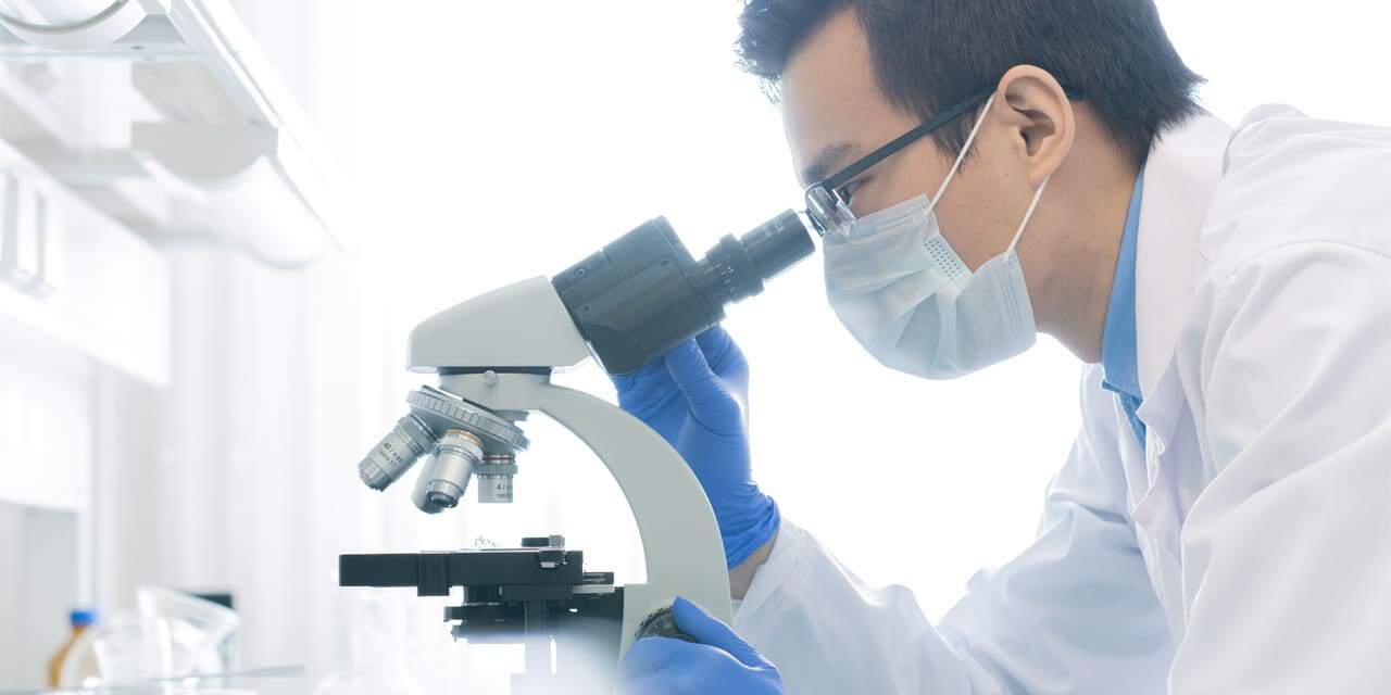
Highlighted in National Catholic Register, there have been three studies published in the last half of 2020 showing how scientists are using preborn remains for “research.”
Published in September 2020, one study focuses on growing “full-thickness human skin” as “the human skin is a significant barrier for protection against pathogen transmission.” It does this by grafting “human full-thickness fetal skin, autologous fetal lymphoid tissues and autologous fetal liver-derived hematopoietic stem cells” on the backs of humanoid mice.
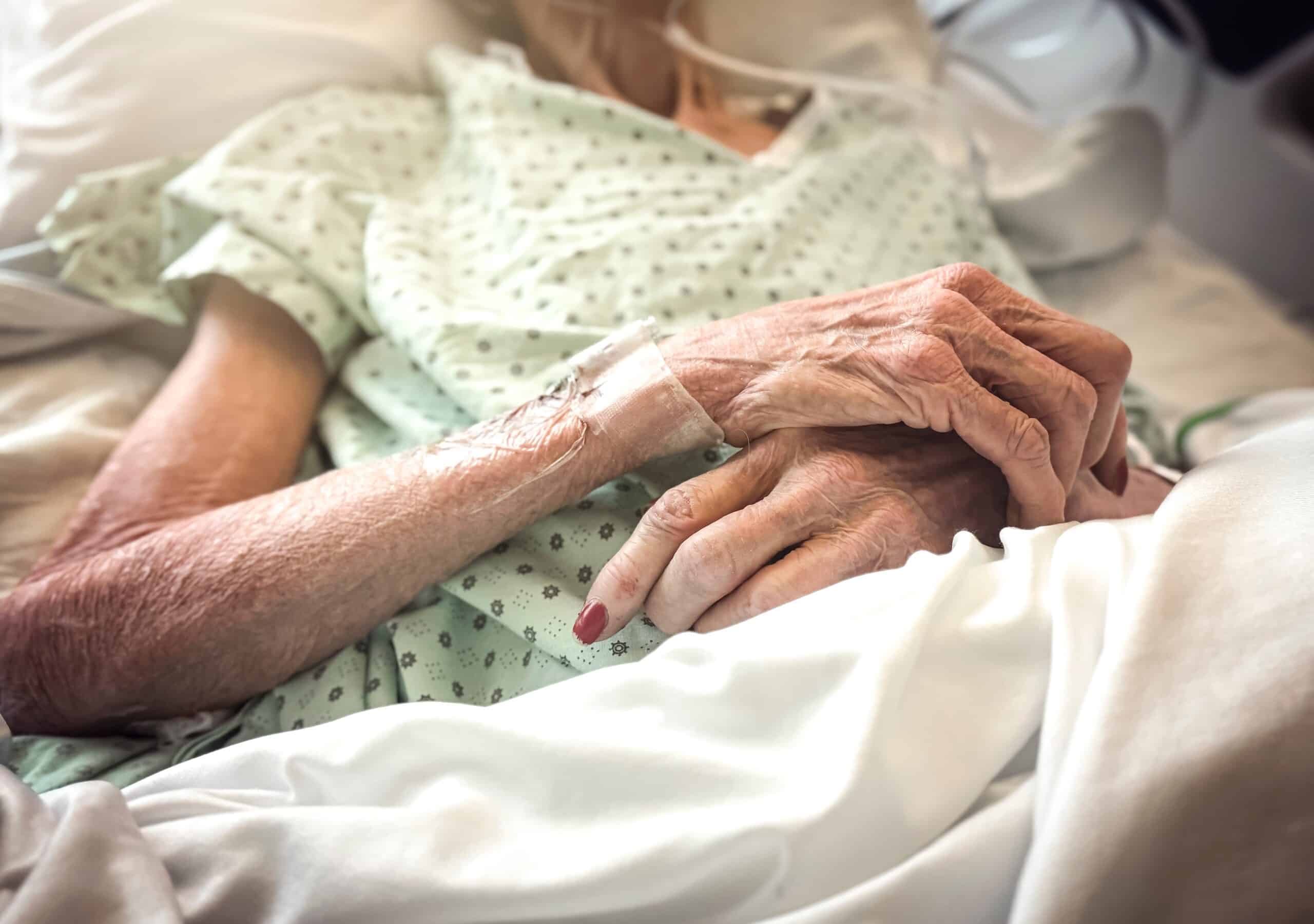
The tissue was taken from the scalp and back of aborted babies between the gestational ages of 18-20 weeks old. At this point in pregnancy, the external sex characteristics are visible on the ultrasound, the mother is feeling the baby move and the baby is preparing for life outside the womb by developing his or her reflexes by sucking their tongue. These babies were literally weeks or days away from viability, which can be as low as 21 weeks.
That might not mean much, but as they say, a picture is worth 1,000 words. This image, published in the research paper, disturbingly shows human hair growing from some of the preborn skin from the scalp on the back of rats, which was done by design to compare. Some of the visible hair is likely the baby’s own, which had just started growing only weeks before the abortion.
The second study also used preborn babies aborted in the second trimester and was designed to determine how the caustic effects of a flame retardant impacts fetal brain development during pregnancy on people of different racial and ethnic backgrounds. Known as polybrominated diphenyl ethers (PBDEs), it’s a chemical that’s found in furniture, electronics and mattresses.
If a mother is exposed to PBDEs, the chemical can transfer to the preborn through the mother’s placenta and the “liver cannot metabolize the chemicals as readily, PBDEs collect in the developing child and continue to build in infancy and childhood, all critical times for the development of the endocrine, immune and neural systems.”
So, in order to determine how this happens abortionists from the University of California San Francisco, a notorious abortion research hub, obtained the consent of 249 women scheduled for a second-trimester abortion in order to dissect the baby’s liver so they could compare the mother’s and child’s levels.
This study is being funded by the U.S. Environmental Protection Agency and the National Institute of Environmental Health Services.
The final study, conducted at Yale University’s Department of Immunology, studied the “development of immunities in newborns” since research has shown that “newborn blood samples show abundant auto-antibodies.”
These babies were once again the victims of a second trimester abortion and had their liver, bone marrow and spleens examined and compared against “blood, bone marrow and stool samples from health adults” to determine the antibody production and gut microbiota.
There’s a saying, just because you can doesn’t mean you should.
These studies are unnecessary and, honestly, evil. Why is an aborted baby scalped and its skin placed on the back of a mouse? What is the purpose, and will this really help anyone?
Medical research has provided humanity with great insight, but it has also been used to commit horrible atrocities in the pursuit of knowledge. The Nazi regime is a great example of what happens when ethics, the sanctity of life and human dignity take a back seat to science.
Photo from Shutterstock


ABOUT THE AUTHOR